Table of Contents
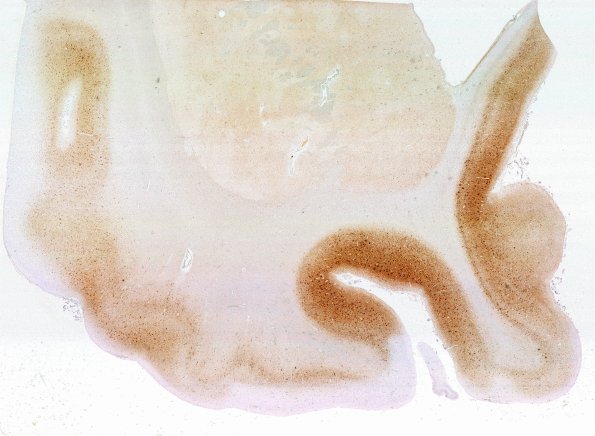
Washington University Experience | NEURODEGENERATION | Alzheimer Disease | Beta Amyloid Plaques | 5G1 AD & CAA L6 PHF WM
The same area shows different cortical and basal ganglia areas stained with PHF. There is a homogeneous pattern involving the neuropil. Compare with 5F1. (PHF IHC)

