Table of Contents
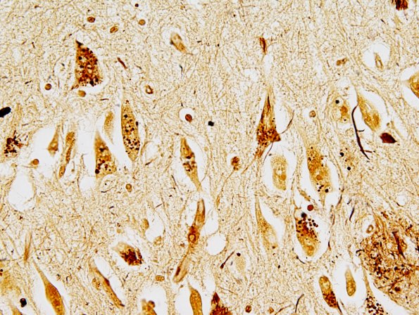
Washington University Experience | NEURODEGENERATION | Alzheimer Disease | Granulovacuolar Degeneration | 2A1 AD N3 Biels 2.jpg
2A1-3 GVD is illustrated as punctate argyrophilic granules in these Bielschowsky silver stained preparations. Notice the neuronal neurofibrillary tangle (arrow, 2A2) and neuritic plaques (arrowheads, 2A2).

