Table of Contents
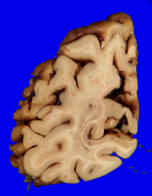
Washington University Experience | NEURODEGENERATION | Alzheimer Disease | Gross Pathology | 5A6 AD (grossly looks like FTD, Case 5)_17
The occipital lobe is also atrophic. ---- Neuro Microscopic Summary: Globally sections of the neocortex and hippocampus showed profound neuronal loss and gliosis in the cortex and amyloid deposits, a number with "cotton-wool"-appearance. An antibody against beta-amyloid (10D5) highlights these amyloid plaques. Neuronal tangles are identified in Ammon's horn on routine H&E stained sections. A Bielschowsky silver stain and an immunostain for paired helical filaments (PHF) highlight scattered neuronal tangles and neuritic plaques in the neocortex as well as the hippocampus; PHF also highlights scattered neuropil threads and intraneuronal pretangles. Immunostains for alpha-synuclein and TDP-43 are negative. ---- Neuro Diagnosis Comment: The patient received a diagnosis of Alzheimer disease neuropathologic change A3B3C3 corresponding to an "high" score in the NIA-AA diagnostic scheme, and reason to believe that Alzheimer disease was responsible for this patient's dementia and terminal course. There were no Lewy bodies or other unusual neuronal inclusions to suggest a concurrent disease process such as Parkinson's disease or diffuse Lewy body disease (also called dementia with Lewy bodies). No compelling evidence for fronto-temporal dementia was identified.

