Table of Contents
Washington University Experience | NEURODEGENERATION | Alzheimer Disease | Synaptic Pathology | 2A1 AD (Case 2)
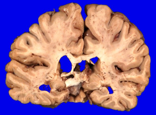
The weight of the unfixed brain was 1185g. There is mild diffuse atrophy of the frontal, parietal and temporal lobes. The hippocampi are small and the amygdalae are atrophic. The vascular disorder was most closely consistent with cavernous angiomas. Diffuse and neuritic amyloid plaques were frequent and neurofibrillary tangles were moderate to frequent. Using the NIA-AA grading scheme this is consistent with Alzheimer disease explaining her dementia. By the NIA-AA scheme she has Alzheimer Disease Neuropathology Change A3B3C3.

