Table of Contents
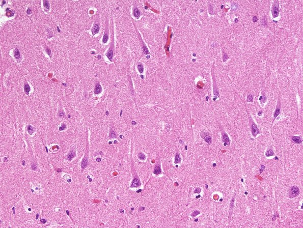
Washington University Experience | NEURODEGENERATION | Argyrophilic Grain Disease (AGD) | 1B2 AGD (Case 1) L23 H&E 2.jpg
1B2-4 Most of the involved temporal lobe and amygdala shows minimal histopathology. Although there is focal neuron loss and astrocytosis, higher magnification shows nothing distinctive to attest to the presence of AGD. (H&E)

