Table of Contents
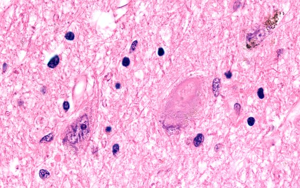
Washington University Experience | NEURODEGENERATION | Corticobasal Degeneration (CBD) | 4C5 CBD (Case 4) N6 H&E 100X 3
A compelling tangle within a substantia nigra neuron. (H&E) ---- Not shown: The microscopic appearance of the cerebellum is unremarkable with a well-preserved complement of Purkinje, granular and dentate neurons. A Bielschowsky stain verifies the absence of neuritic plaques and neurofibrillary tangles, and alpha-synuclein fails to identify any Lewy bodies, either nigral or cortical. Neurofilament however highlights numerous scattered swollen/chromatolytic neurons in the cingulate gyrus, as well as frontal cortex and the basal ganglia. No Lewy bodies are identified. ---- Neuro Final Diagnosis: Corticobasal Degeneration (see Comment) ---- Neuro Diagnosis Comment: The neurodegenerative process in this patient is characterized by several histopathologic findings including neurofilament positive swollen neurons within the cerebral cortices, prominent neuronal drop-out within the basal ganglia (specifically the globus pallidus) and marked diminution of neurons in the substantia nigra. Taken together, these features are characteristic of Corticobasal Degeneration, a syndrome that was suggested by this patient's clinical history. The absence of classical neurofibrillary tangles, the anatomic distribution of pathology and absence of neuritic plaques eliminated several diagnoses, specifically progressive supranuclear palsy and Alzheimer's disease. The absence of Lewy bodies within the nigra and cortex additionally excludes Parkinson's disease and diffuse Lewy body disease. Finally, this case lacked the tau-positive glial cytoplasmic inclusions characteristic of multi-system atrophy.

