Table of Contents
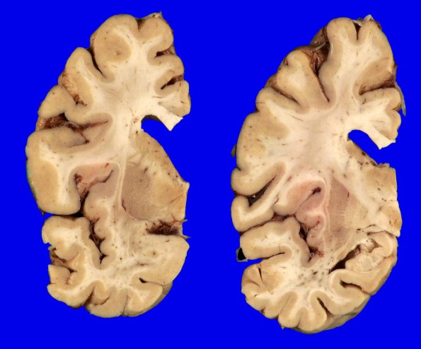
Washington University Experience | NEURODEGENERATION | Hippocampal Sclerosis of Aging | 3A2 HScl (Case 3) Gross_1
There was mild to moderate atrophy of the frontal, temporal, and parietal lobes. Coronal slicing revealed mild to moderate dilatation of the lateral ventricle with rounding of the angles. The hippocampus was atrophic. The substantia nigra and locus coeruleus were normally pigmented.

