Table of Contents
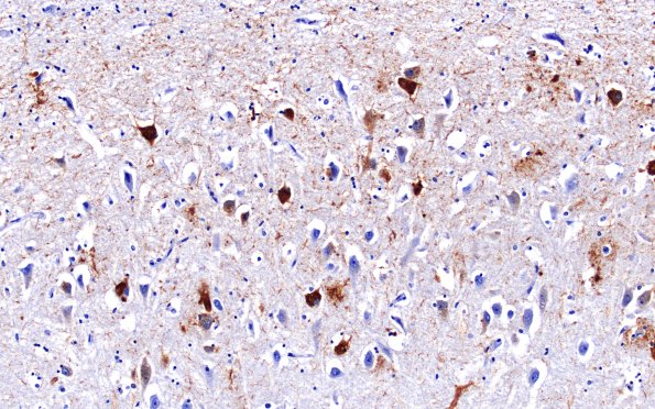
Washington University Experience | NEURODEGENERATION | Hippocampal Sclerosis of Aging | 5D2 Hippocampal Sclerosis (Case 5) N3 PHF 20X
The hippocampal formation does show neuritic plaques and neurofibrillary tangles in the CA1 area in particular and extending into the subiculum and adjacent entorhinal cortex. (PHF1 IHC) ---- Not shown: Multiple sections of frontal, parietal, temporal and occipital neocortices show a relatively well maintained complement of neurons generally without marked astrocytic and microglial proliferation or parenchymal perivascular inflammatory cells. Pick bodies, cortical Lewy bodies, neoplasia, malformation and ballooned cells are not demonstrated in H&E, beta-amyloid, and phospho-tau stained material of the neocortex. There are scattered senile plaques demonstrable in H&E stained neocortex confirmed by thioflavin-S, Bielschowsky, and immunostains. Neuron loss, astrocytosis, scattered plaques and tangles are found in the amygdala. The appearance of underlying digitate and deeper white matter shows mild focal pallor which, although involved by modest arteriolosclerosis, shows very little ischemic pathology, microthrombi or amyloid deposition. .

