Table of Contents
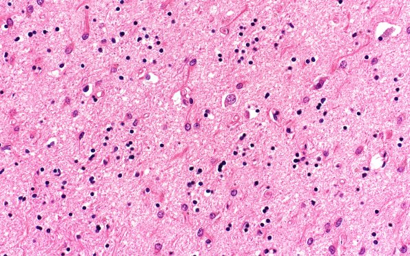
Washington University Experience | NEURODEGENERATION | Huntington Disease | 11B3 Huntington Dz (Case 11) H&E 40X
Microscopic examination of the basal ganglia shows marked neuron loss and astrocytosis involving the caudate, putamen, and to a lesser extent, the globus pallidus. There is marked atrophy and contraction of these areas with prominent gliosis. Occasional larger neurons are still present, however the population of small neurons is markedly diminished. ---- Not shown: Multiple sections of the cerebral cortices show a thinned cerebral cortex and normal-appearing white matter. The density of the cortical neuronal population appears within normal limits and there is no significant gliosis. Although a few macrophages in the substantia nigra contain pigment, neither Lewy bodies nor astrocytosis are seen. Sections of the pons and medulla are unremarkable; specifically, the locus ceruleus in the pons has a well-preserved population of pigmented neurons. ---- Neuro Diagnosis Comment: Neuropathologic examination reveals the histologic findings diagnostic of Huntington's disease. These include marked neuron loss and astrocytosis in the caudate, putamen, and globus pallidus. The patient has no neuropathologic evidence of any additional neurodegenerative diseases, specifically no evidence of Lewy body Parkinson's disease. Despite prominent parkinsonian features of his clinical illness, the patient's morphologic findings are those of Huntington's disease, consistent with his genetic phenotype.

