Table of Contents
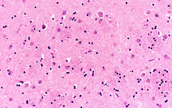
Washington University Experience | NEURODEGENERATION | Huntington Disease | 12B Huntington Dz (Case 12) 40X
Microscopic examination of the deep grey structures including the basal ganglia show moderate neuronal loss and astrocytosis of the caudate, putamen and, to a lesser extent, the globus pallidus. ---- Not shown: Multiple sections of neocortex including sections from the frontal, parietal, temporal and occipital lobes show mild patchy subpial gliosis and patchy astrocytosis in the cortex. Scattered microinfarcts were identified most prominently involving the cerebral white matter and the structures in the posterior fossa. In some regions these are associated with tiny perivascular hemorrhages and clear spaces. The latter and the white matter distribution suggests the possibility of fat embolism. Alternatively, DIC may have contributed. In either case, sepsis was the most likely etiology. No viral inclusions were identified, and there was no evidence of meningitis or encephalitis.

