Table of Contents
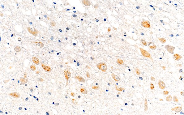
Washington University Experience | NEURODEGENERATION | Huntington Disease | 15E Huntington's Dz (Case 15) Midbrain L9 1C2 40X
Scattered neurons had inclusions in the region of the midbrain 3rd nerve motor nucleus. Neurons with lipofuscin alone did not have this pattern. (1C2 IHC) ---- Not shown: Immunohistochemistry revealed no aggregates reactive for amyloid-beta peptide, phosphorylated synuclein, TDP-43, or tau. ---- Neuro Diagnosis Comment: Macroscopy shows atrophy of the caudate nucleus consistent with Grade 2 (range: 0, normal to 4, severe) in the Huntington's Disease Neuropathological Grading Scheme of Vonsattel et al. (PMID: 9596408). Histological slides show severe neuronal loss and gliosis in the caudate nucleus and, to a lesser degree, in the putamen. There is also very mild neuronal loss in neocortical areas, including the frontal, temporal, and parietal lobes. 1C2 immunohistochemistry detects polyglutamine aggregates in the form of neuronal intranuclear inclusions that are the histological hallmark of Huntington's disease. Previous molecular genetic analysis confirmed the polyglutamine expansion in the huntingtin gene.

