Table of Contents
Washington University Experience | NEURODEGENERATION | Huntington Disease | 16A1 Huntington's Dz (Case 16) 1
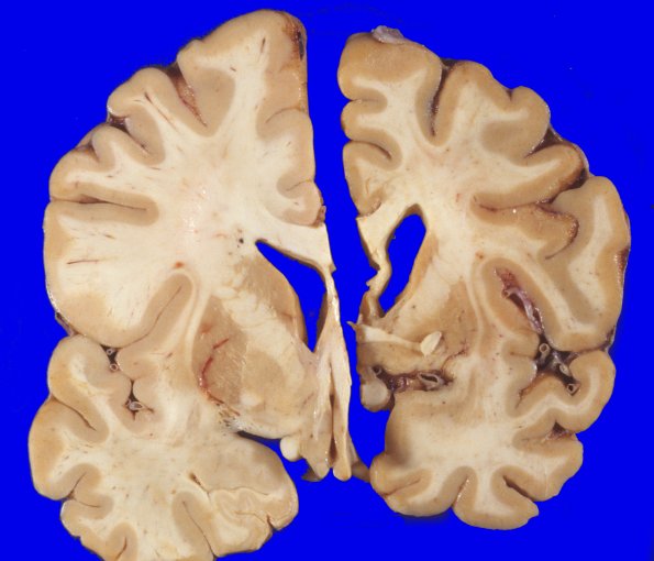
16A1-3 Coronal sections through a control cerebral hemisphere (left hemibrain) compared to this HD case (right hemibrain) demonstrate a mild degree of cortical atrophy involving the frontal and temporal lobes. The angle of the lateral ventricle is blunted. The basal ganglia are markedly abnormal. The caudate nucleus is reduced to a 1-2 mm. discolored strip which does not protrude into the lateral ventricle. The putamen and globus pallidus were less markedly atrophic but were also discolored.

