Table of Contents
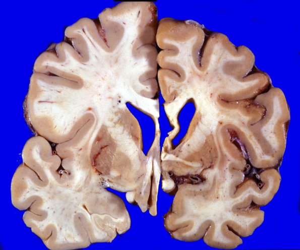
Washington University Experience | NEURODEGENERATION | Huntington Disease | 16A3 Huntington's Dz (L) vs Normal Brain (R) Case 16
Coronal sections through a control cerebral hemisphere (left hemibrain) compared to this HD case (right hemibrain) demonstrate a mild degree of cortical atrophy involving the frontal and temporal lobes. The angle of the lateral ventricle is blunted. The basal ganglia are markedly abnormal. The caudate nucleus is reduced to a 1-2 mm. discolored strip which does not protrude into the lateral ventricle. The putamen and globus pallidus were less markedly atrophic but were also discolored. ---- Not shown: All areas of cerebral cortex showed a relatively well-preserved complement of cortical neurons. In some places there is an apparent slight decrease in neuronal number which was, however, not accompanied by a focal increase in number of astrocytes. Neuron loss appears to preferentially spare large neurons. In some areas (particularly anterior caudate) appreciable numbers of small neurons remain, which extend into the nucleus accumbens. The remaining neurons show no appreciable increase in lipopigment or cytoplasmic inclusions with H&E stain. The bundles of myelinated axons within the putamen and globus pallidus appear more densely aggregated and atrophic presumably as a result of loss and collapse of intervening neuropil and neurons.

