Table of Contents
Washington University Experience | NEURODEGENERATION | Huntington Disease | 2A3 Huntington's Dz (Case 2)_13
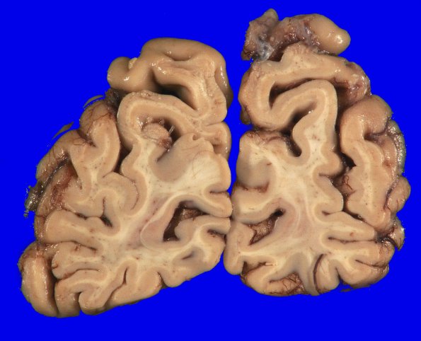
Coronal sections of the cerebral hemispheres reveal a global atrophic pattern with widening of sulci, again most pronounced in the insular, temporal and occipital areas. The corpus callosum is somewhat thinned, and the white matter overall has an atrophic appearance. The lateral ventricles are blunted and dilated, consistent with an ex vacuo appearance and basal ganglia atrophy. The caudate nucleus of the basal ganglia retains a convex appearance.

