Table of Contents
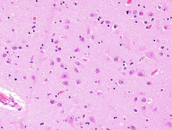
Washington University Experience | NEURODEGENERATION | Huntington Disease | 2B Huntington's Dz (Case 2) N3 H&E 21.jpg
Hematoxylin and eosin stained sections of frontal, temporal, parietal, and occipital lobes show mild subpial gliosis and minimal cortical neuron loss or parenchymal astrocytosis.

