Table of Contents
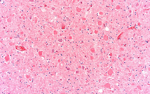
Washington University Experience | NEURODEGENERATION | Infantile Neuroaxonal Dystrophy (INAD) | 1D1 INAD (Seitelberger Dz, Case 1) E1 Thal 20X 2
1D1,2 These sections show several areas of the thalamus/basal ganglia with prominent dystrophic axons, often adjacent to neuronal cell bodies (arrow, 1D2). They appear more plentiful in the caudate nucleus than in the lentiform nuclei, and in the former are associated with astrocytosis and neuron loss. Extracellular, parenchymal iron and calcium positive pigment deposition was prominent in the medial segment of the globus pallidus accompanied by neuron loss and astrocytosis (not shown). (H&E)

