Table of Contents
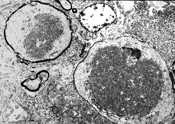
Washington University Experience | NEURODEGENERATION | Infantile Neuroaxonal Dystrophy (INAD) | 2C5 INAD (Seitelberger Dz, Case 2) EM2
2C5,6 Ultrastructural examination of swollen dystrophic axons showed they often maintained a thin rim of attenuated myelin at their margins. They contained large numbers of tubulovesicular profiles, often arranged in a paracrystalline lattice, membranous lamellae and clear clefts, tubular rings, and a variety of mitochondrial alterations including glycogen deposition as granular and dense bodies. Such ultrastructural alterations are distinctive and characteristic of neuroaxonal dystrophy. Ultrastructural appearance of dystrophic axons is often dominated by dilated thinly myelinated axons containing aggregates of tubulovesicular elements. (electron micrographs)

