Table of Contents
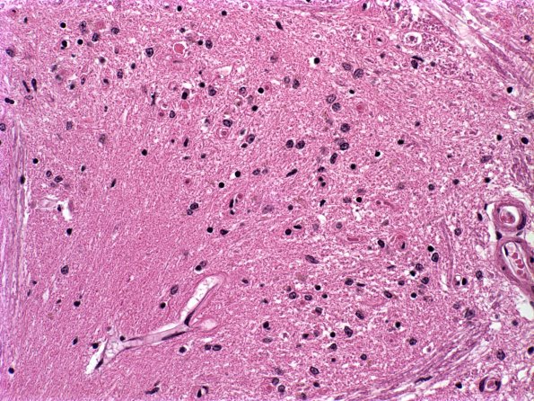
Washington University Experience | NEURODEGENERATION | Multiple Systems Atrophy (MSA) | 17C3 MSA (Case 17) ION a
Higher magnification of image #17C2. (H&E) ---- Not shown: The substantia nigra and locus ceruleus are normally pigmented. The aqueduct and the fourth ventricle have a normal configuration and are lined by a smooth, glistening ependyma. Radial sections of the cerebellum show normal foliar architecture. The cortex, white matter and dentate nuclei are grossly unremarkable. Sections of the spinal cord are grossly unremarkable. The pons shows relatively normal numbers and pigmentation of the neurons of the locus ceruleus. ---- Neuro Final Diagnoses: Multiple Systems Atrophy (MSA); Infarcts, right pons, remote ---- Neuro Diagnosis Comment: This is an interesting and somewhat atypical case. Although the glial cytoplasmic inclusions diagnostic for MSA are unequivocally present in this case, their distribution and the focal nature of the pathologic findings is somewhat unusual for typical MSA cases. Specifically, the involvement of the cerebellum and inferior olivary nuclei typically are accompanied by substantial neuron loss in the pons which is not prominent in this case, although large numbers of GCI are present in the pons. The loss of Purkinje cells is modest and patchy, although the presence of large numbers of axonal swellings involving residual Purkinje cells may effectively disconnect cerebellar cortical neurons from the dentate nucleus, even if its neurons are spared. There is no evidence of striatonigral or Shy-Drager patterns of injury.

