Table of Contents
Washington University Experience | NEURODEGENERATION | Neuronal Nuclear Inclusion Disease (NIID) | 2A2 NIID & Alzheimer Dz (NIID, Case 2) A10
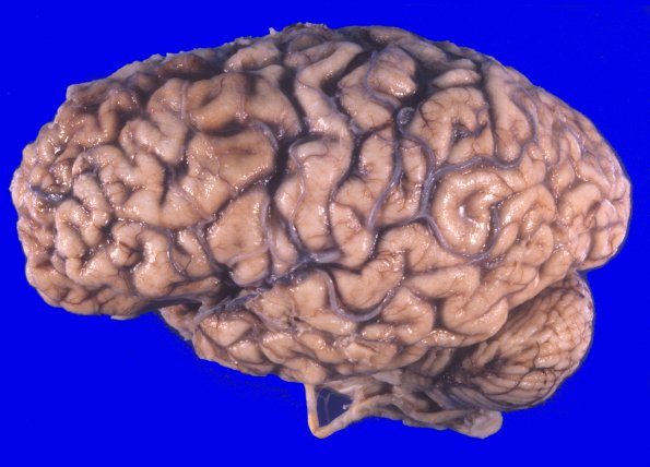
The extent of cortical atrophy, marked in the frontal and temporal lobes, is well shown by this lateral image with gaping sulci and narrowed cortical gyri. There are no external signs of softening, hemorrhage, herniation, or discoloration. The cerebellar hemispheres are full and equal in size.

