Table of Contents
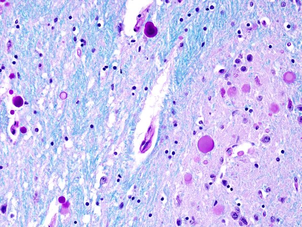
Washington University Experience | NEURODEGENERATION | Polyglucosan Body Disease (PGBD) | 3B4 PGB (Case 3) DHorn LFB-PAS 1
3B4,5 The populations of PGB differ qualitatively in LFB-PAS stained dorsal column and adjacent dorsal horns; specifically, PGB in dorsal column white matter (images 3B1-3) have a normal dark purple appearance and are typically found next to the pia at the margins of the cord and around individual parenchymal vessels. Adjacent areas of the dorsal horn show very large numbers of light pink stained PGB which differ qualitatively and clearly from those in adjacent white matter tracts. We failed to identify clear-cut neurons containing these inclusions in the dorsal horn. (LFB-PAS)

