Table of Contents
Washington University Experience | NEURODEGENERATION | Polyglucosan Body Disease (PGBD) | 3G3 PGB (Case 3) SC EM 019 - Copy
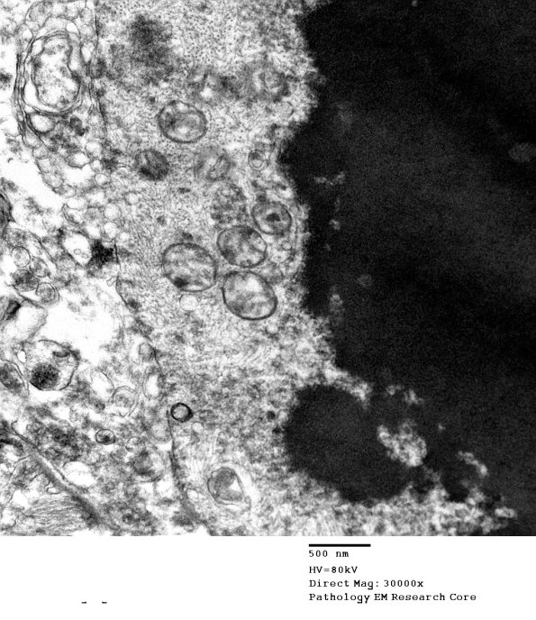
3G2,3 This unusual structure is reminiscent of one of the patterns seen in neuroaxonal dystrophy in humans and animals. (electron micrograph) ---- Diagnosis: Marked increase in polyglucosan bodies in white and gray matter, particularly rostral spinal cord. ---- Not shown: Sections of the cerebral cortex show a well-preserved complement of neurons, and only a mild increase in numbers of cortical astrocytes. Sections of the hippocampus fail to identify significant numbers of senile plaques, neurofibrillary tangles, cortical Lewy bodies, Hirano bodies or granulovacuolar degeneration. There are questionably increased numbers of corpora amylacea in the subpial areas including perivascular areas which abut the Virchow-Robin spaces but not obviously a marked increase in numbers in the cortical and white matter. Sections of the basal ganglia are remarkable for substantial increase in corpora amylacea and rare neurons undergoing eosinophilic neuronal necrosis. Sections of the midbrain and pons are remarkable for a few eosinophilic neurons although the neuronal population is well maintained in motor nuclei and pigmented neuronal nuclei.

