Table of Contents
Washington University Experience | NEURODEGENERATION | Progressive Supranuclear Palsy (PSP) | 16A3 Progressive Supranuclear Palsy (Case 16) 3
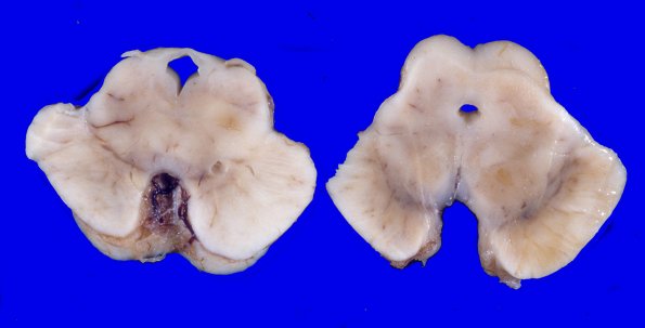
The substantia nigra are patchy pale and there is a hint of atrophy of the midbrain tectum/tegmentum. ---- There are no slides available from the autopsy but I will report the findings. Sections show neuron loss and gliosis in the basal ganglia, thalamus, subthalamic nucleus, dentate nucleus, and substantia nigra. These areas also show neurons with neurofibrillary tangles and occasional neuritic and non-neuritic plaques by Bielschowsky silver stain. In addition, the substantia nigra showed free pigment in the neuropil and pigment laden histiocytes. Oculomotor nuclei and neurons in the periaqueductal grey matter show neurofibrillary tangles. Neurofibrillary tangles were also present in the loci cerulei. There are several neurofibrillary tangles, and occasional non-neuritic plaques in the hippocampi on Bielschowsky silver stains. ---- Final Neuropathologic Diagnosis: Progressive supranuclear palsy ----Comment: Neuropathologic examination of the brain showed neuron loss, gliosis, neurofibrillary tangles and plaques in basal ganglia, thalamus, subthalamic nuclei, dentate nuclei, and substantia nigra. Prominent atrophy of the frontoparietal areas or ballooned neurons in cortical grey matter, to indicate cortical basal ganglionic degeneration, are not present. The frequency of neuritic and non-neuritic plaques are not compelling for Alzheimer's disease. No Lewy bodies are present in the substantia nigra or locus cerulei.

