Table of Contents
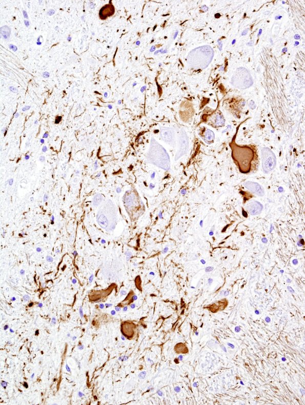
Washington University Experience | NEURODEGENERATION | Progressive Supranuclear Palsy (PSP) | 1C18 PSP (Case 1) N7 Medulla PHF1 3.jpg
The ION and adjacent raphe nucleus are prominently stained for pTau. (PHF1 IHC) ---- Not shown: There was no evidence of amyloid plaques or cerebral amyloid angiopathy in the frontal lobe, temporal lobe or hippocampus. Basophilic globose inclusions were identified in neurons of the striatum and subthalamic nuclei. PHF-1 IHC shows abundant tau-positive intraneuronal globose inclusions and neuropil threads in the subthalamic nuclei, and frequent neuronal and glial tau-positive inclusions in the thalamus (inferomedial>superolateral). IHC for phosphorylated alpha-synuclein, performed on the amygdala, midbrain, and medulla, shows no evidence of Lewy bodies or other aggregates of phosphorylated alpha-synuclein. Sections of the cerebellum show relatively well-preserved cortical layers, albeit with occasional Purkinje cell neuroaxonal swellings (‘torpedos’). The dentate nucleus shows no overt neuronal loss or inclusions, but grumose degeneration of axon terminals associated with dentate nucleus neurons is appreciated. Ballooned neurons, though identified within the amygdala, were not detected in sufficient numbers in the cortex to suggest another diagnosis such as corticobasal degeneration. No deposits of amyloid-beta peptide or aggregates of alpha-synuclein were detected, excluding diagnoses of Alzheimer disease, Parkinson disease, and multiple systems atrophy (MSA). ---- Final Neuropathologic Diagnoses: Progressive supranuclear palsy (PSP); Intraventricular hemorrhage.

