Table of Contents
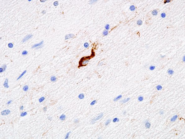
Washington University Experience | NEURODEGENERATION | Progressive Supranuclear Palsy (PSP) | 2D4 PSP (Case 2) Pons, Coiled body, PHF-6
A coiled body involving an oligodendrocyte in the basis pontis. (PHF1 IHC) ---- Not shown: The neocortices showed mild neuronal loss and gliosis. Frequent diffuse and few cored beta-amyloid plaques are seen. However, there are numerous tau-positive neurofibrillary pre-tangles, tangle-like inclusions, tufted astrocytes, and coiled bodies in oligodendrocytes in the gray matter. There are no cortical Lewy bodies. There is neuronal loss and gliosis, some swollen achromatic neurons, focally numerous tau-positive neuronal pre-tangles, tangles, neuritic plaques, tufted astrocytes, and coiled bodies in the amygdala. In the entorhinal cortex, there is severe neuronal loss, microvacuolation in superficial laminae, focally numerous tangles and neuritic plaques. Lewy bodies are not detected. There is some loss of neurons and gliosis in the subthalamic nucleus. There are focally numerous pTau immunoreactive tangles in both the thalamus and subthalamic nuclei. The substantia nigra and locus ceruleus shows moderate to severe loss of neurons, extracellular pigment, and gliosis. Neuronal loss is also discerned in the oculomotor nucleus. No Lewy bodies are present. There are numerous neuronal tau-positive cytoplasmic neurofibrillary pre-tangles and scattered astrocytic inclusions. The ION contains numerous tangles. ---- Neuro Final Diagnosis: Progressive supranuclear palsy; Alzheimer disease neuropathologic change (see comment) ---- Neuro Diagnosis Comment: This case shows the stereotypic features of progressive supranuclear palsy (PSP), also known as Steele-Richardson-Olszewski syndrome: degeneration including widespread neuronal loss, gliosis, tau-positive neuronal and glial cytoplasmic inclusions, especially tufted astrocytes, in cortical and subcortical nuclei. Histological slides of representative neocortical areas in this case show frequent beta-amyloid plaques, but only few neuritic plaques are present. The neurofibrillary tangles are most numerous in the medial temporal lobe. The distribution of beta-amyloid plaques, sparse neuritic plaques, and tangles are consistent with stage IV in the Braak and Braak stageing of tangles and amyloid stage C. These findings are sufficient to meet the neuropathologic criteria for the diagnosis of Alzheimer's disease (AD) according to the criteria of Khachaturian, but not CERAD, or NIA-Reagan Institute.

