Table of Contents
Washington University Experience | NEURODEGENERATION | Wilson Disease | 1A2 Wilson Dz (Case 1) 3A
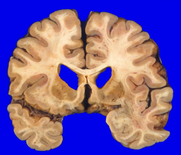
Coronal sections of the cerebral hemispheres show the caudate nuclei are yellowish-orange and atrophic. The caudate heads are concave, i.e.. they do not bulge into the lateral ventricles. The putamina are soft and mildly discolored with focal cavitation (1A2). The ventricles are dilated and blunted which largely reflects the loss of corpus striatum mass. The cerebral cortex appears largely intact with little atrophy.

