Table of Contents
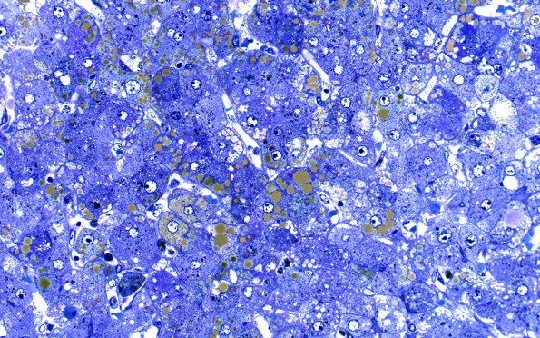
Washington University Experience | NEURODEGENERATION | Wilson Disease | 4A2 Liver Copper (Case 4) Plastic 40X
This plastic section shows loss of acinar architecture. There is both canalicular and ductal cholestasis. The hepatocytes within the regenerative nodules show prominent ballooning degeneration, and micro- and macrosteatosis. (One micron thick toluidine blue stained plastic section) ---- Not shown: Sections show a diffuse macro and micronodular cirrhosis. The fibrous bands separating regenerative nodules contain prominent bile ducts and ductules. The trichrome and reticulin stains demonstrate bands of collagen and the loss of acinar architecture, respectively. Given the turnover of hepatocytes in the regenerative nodules, it is not uncommon for the copper stain to demonstrate a relatively modest degree of copper accumulation.

