Table of Contents
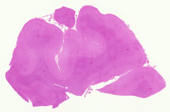
Washington University Experience | PRION DISEASES | Prion Diseases | 11A2 CJD (Case 11) H&E occipital cortex WM
11A2-4 H&E stained sections of the cerebrum show diffuse cortical spongiform changes, neuronal loss, and gliosis; most pronounced in the occipital lobe compared to the frontal and temporal lobes. The basal ganglia and thalamus show some mild spongiform change and gliosis. The molecular cell layer of the cerebellum is shows only mild vacuolation. The remainder of the cerebellum is well-preserved. There are no 'florid' or 'kuru-like' plaques seen in the material examined.

