Table of Contents
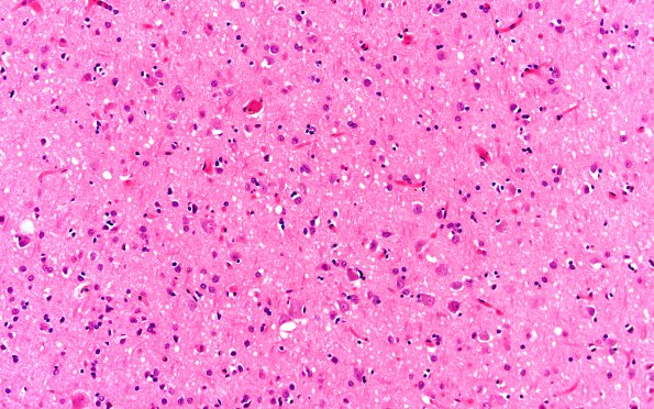
Washington University Experience | PRION DISEASES | Prion Diseases | 13A2 CJD (Case 13) H&E N1 20X
Sections from the selected neocortical regions, including frontal, temporal, parietal and occipital (shown) lobes, all show pathology characterized by numerous 5-25 micron vacuoles in neurons and neuropil, consistent with spongiform encephalopathy.

