Table of Contents
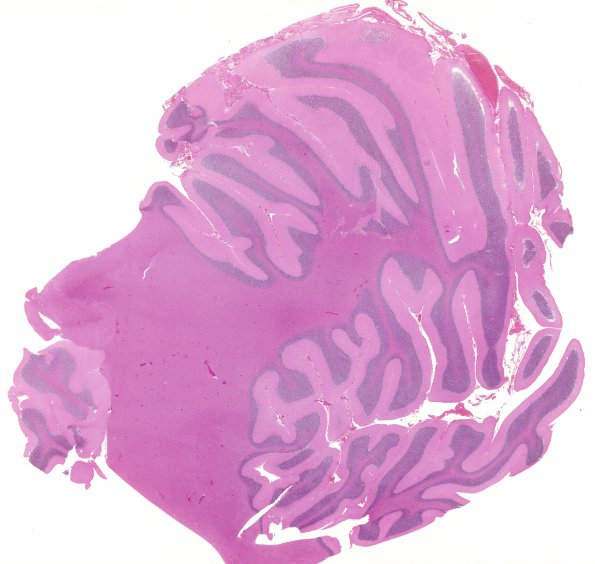
Washington University Experience | PRION DISEASES | Prion Diseases | 18B1 CJD (Case 18 H&E WM
18B1-3 Sections of the frontal, temporal, and occipital cortices, as well as the thalamus show diffuse but generally mild spongiform change, neuron loss, and astrogliosis, most prominent in the cerebellum. The vacuoles are small and round, and involving the neuropil of grey, predominantly, and white matter. The cerebellum shows spongiform change in the molecular layer as well as vacuolation involving the interfoliar white matter. Significantly there are no “florid” plaques present that would suggest a diagnosis of variant CJD.

