Table of Contents
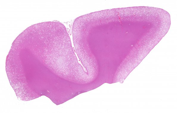
Washington University Experience | PRION DISEASES | Prion Diseases | 1C1 CJD (Case 1) Temporal cortex H&E whole mount 1A
1C1-3 Sections of the frontal, temporal (shown), and occipital cortices show spongiform change, neuronal loss, and prominent astrogliosis. The vacuoles show size variation; larger vacuoles appear to represent multiple merged small vacuoles or loss of parenchyma with increased extracellular space. The distribution of vacuoles within each area is fairly uniform, but the severity of vacuolation, neuronal loss and gliosis is variable within the sampled sections of cortex, decreasing in severity from occipital to frontal to temporal lobes.

