Table of Contents
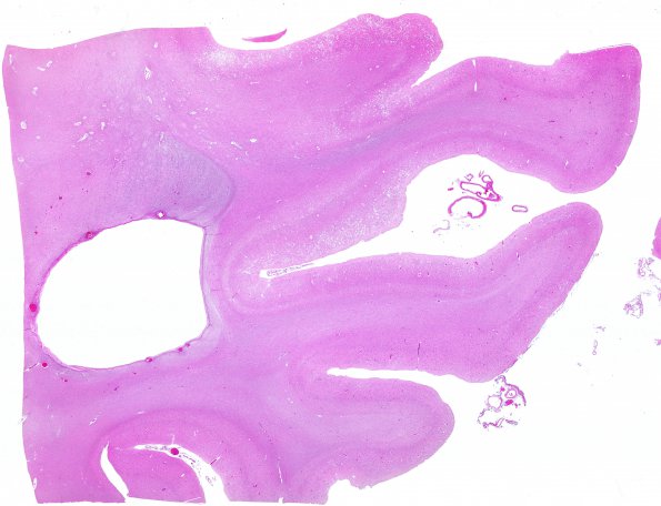
Washington University Experience | PRION DISEASES | Prion Diseases | 20A1 CJD (Case 20) N5 H&E WM
At autopsy the weight of the brain was not weighed for fear of contamination. The external brain surface was unremarkable. Coronal sections through the cerebral hemispheres demonstrate a normal cortical ribbon throughout and no white matter abnormalities. All brain structures were grossly normal. ---- 20A1,2 Sections of the grey matter in the cerebral hemispheres, basal ganglion, thalamus and molecular layer of the cerebellum show spongiform encephalopathy. In these regions there is vacuolation of the neuropil and occasional intra-neuronal vacuoles. The vacuoles are round and regular and range in size from approximately 5-25 um in diameter. Also, there is marked neuron loss and focally severe gliosis.

