Table of Contents
Washington University Experience | PRION DISEASES | Prion Diseases | 25A1 CJD (Case 25) A
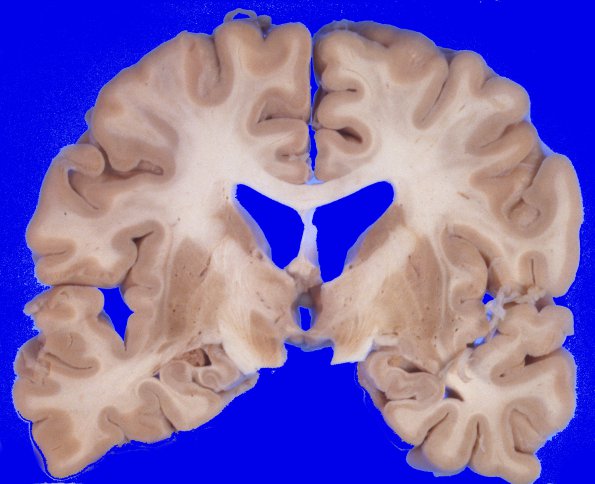
At autopsy brain weight was not reported. The external and cut section appearance showed a degree of atrophy and ventricular dilatation. The cortex is of average and uniform thickness throughout. ---- Sections showed spongiform change involving cerebral cortex, midbrain and corpus striatum. This is accompanied by marked astrocytosis and variable neuronal loss. The gliosis and neuronal loss are most severe in the corpus striatum. Occasional "Kuru" plaques are identified in H&E stained sections. Silver stain of the hippocampus shows rare intraganglionic tangles and neuritic plaques. Sections of cerebellum and brainstem show astrocytosis but without the striking spongiform change. ---- Diagnosis: Spongiform encephalopathy, consistent with Jakob-Creutzfeld disease.

