Table of Contents
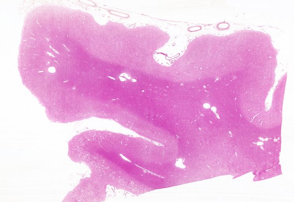
Washington University Experience | PRION DISEASES | Prion Diseases | 26A1 CJD (Case 26) H&E WM
At autopsy the brain was not weighed for fear of contamination. However, the gyri and sulci are described as narrowed and the sulci are greatly deepened. Serial coronal sections of the cerebral hemispheres at 1 cm intervals reveal distinct demarcation between gray and white matter. The cortex is narrowed throughout. The caudate nucleus appears smaller than normal. The ventricles are enlarged but are lined by smooth, glistening ependyma. ---- 26A1-2 Sections show a triad of histopathologic changes: neuronal cell depopulation, dense astrocytic gliosis and a patchy spongiform change. The histologic appearance of the spongiform change is characterized by many small, usually round or oval vacuoles in the gray matter. The spongiform change and neuronal dropout are very prominent in sections of cortex from the temporal lobe. Sections of frontal and occipital cortex and white matter show extreme neuronal loss, white matter pallor and gliosis. Sections of cerebellum show a decreased number of granule cells as well as a white matter gliosis. No additional processes were established; however, the pathology appeared more chronic than several weeks.

