Table of Contents
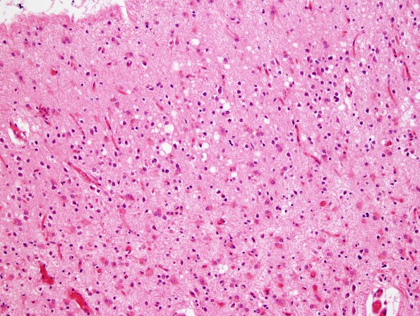
Washington University Experience | PRION DISEASES | Prion Diseases | 2D4 CJD (Case 2) H&E 4
Higher magnification images of the neocortex in multiple lobes shows marked neuronal loss, and extensive astrocytosis with a limited degree of residual spongiform vacuolation. The neuron loss and astrocytosis clearly is transcortical. Adjacent superficial white matter is gliotic. ---- Comment: Tissues sent to the NPDPSC showed granular immunoreactivity with 3F4, the monoclonal antibody to the prion protein. This finding establishes the diagnosis of prion disease, likely Creutzfeldt-Jakob disease.

