Table of Contents
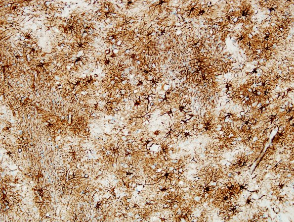
Washington University Experience | PRION DISEASES | Prion Diseases | 3B4 CJD (Case 3) GFAP Basal Ganglia
Astrocytosis is further highlighted by GFAP immunohistochemistry of this basal ganglia section. (GFAP IHC). ---- Not shown: The frontal and occipital lobe cerebral cortices show minimal spongiform change, astrocytosis and neuron loss. There is no evidence of significant numbers of neurofibrillary tangles, Pick bodies or ballooned cells. The appearance of underlying deeper white matter shows mild focal pallor and focally increased numbers of astrocytes. The microscopic appearance of the cerebellum shows minimal vacuolation of the molecular layer. ---- The immunoblot at the NPDPSC reveals the presence of abnormal protease resistant prion PrP 27-30, consistent with the diagnosis of Creutzfeldt-Jakob disease.

