Table of Contents
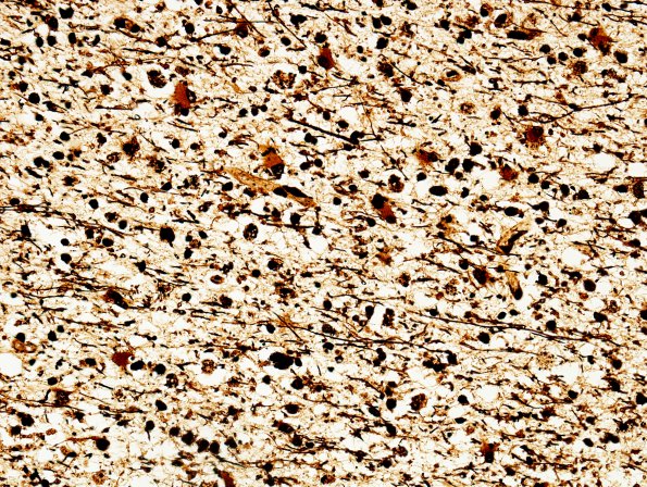
Washington University Experience | PRION DISEASES | Prion Diseases | 4E3 CJD, 5 yr Hx proven PrP (Case 4) CJD, Biels 3
A Bielschowsky silver stain for axons shows no plaques or neurofibrillary tangles but a substantial loss of axons. ---- Not shown: Prion immunohistochemistry reveals diffuse granular staining throughout the cortex and apparent intensely stained prion plaques in the cortex. A similar pattern of synaptic staining is found throughout all the cortical and subcortical nuclei sampled. There is some loss of Purkinje and granule cells, but the cerebellum is relatively well populated. The anti-prion protein antibody (3F4) highlights multiple immunoreactive deposits throughout the cerebellar cortex.

