Table of Contents
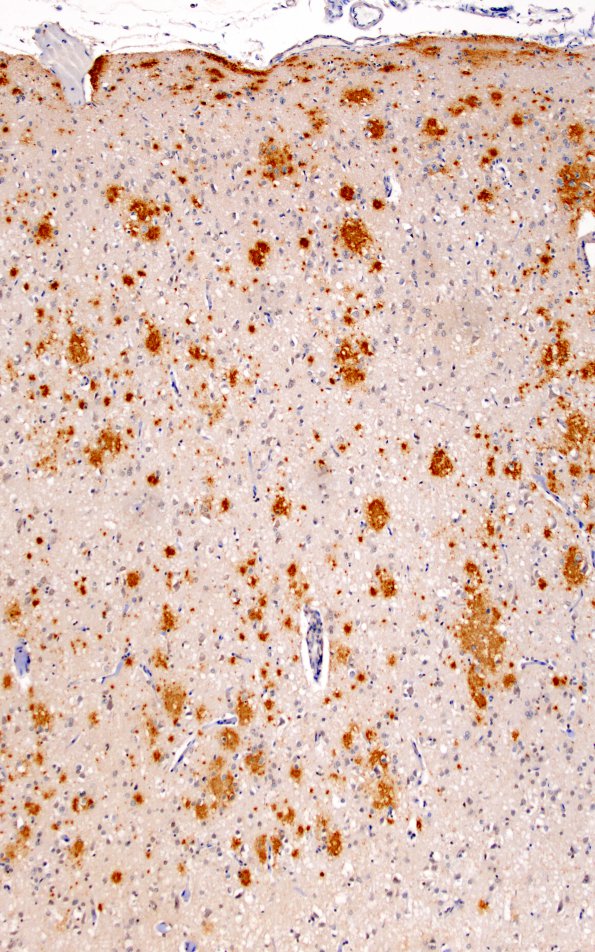
Washington University Experience | PRION DISEASES | Prion Diseases | 9A3 CJD, (Case 9) BAmyloid 10X N4 (HC)
In addition to spongiform changes, Alzheimer's disease like-changes were noted. The frontal cortex shows extensive beta-amyloid plaque deposition by immunohistochemistry and Bielschowsky stain. Bielschowsky stain also shows scattered neurofibrillary tangles and a few neuritic plaques in the cortex. TDP-43 staining is negative in the frontal cortex. Extensive beta-amyloid plaque deposition is also seen in Ammon's horn, the subiculum, and parahippocampal gyrus. There are scattered neurons with granulovacuolar degeneration and neurofibrillary tangles seen in these regions.

