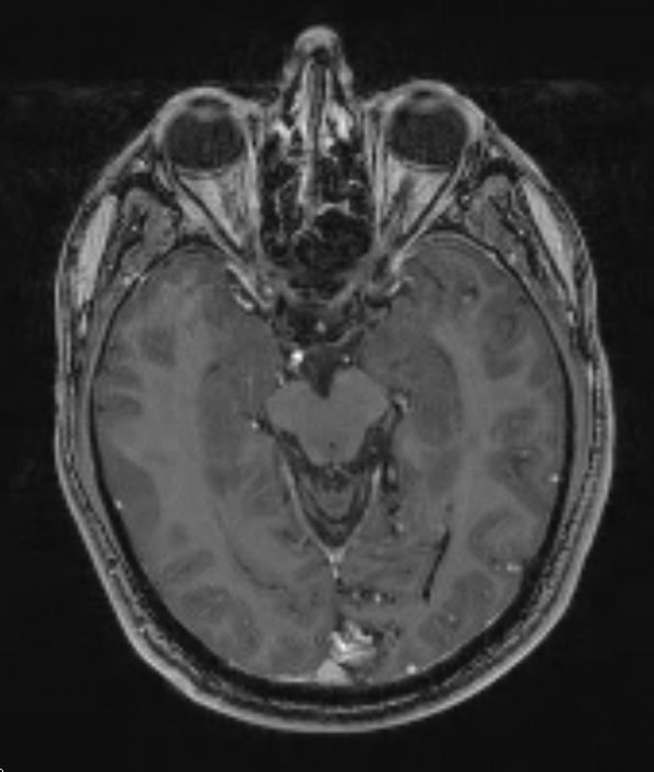
Case 18 History ---- The patient is a 42-year-old man with focal occipital region seizures caused by a left occipital AVM. Surgical resection of the AVM appeared complete as confirmed by an intraoperative indocyanine green angiogram. ---- 18A1,2 This occipital lobe AVM is shown in a T-1 weighted image (18A1) and in an angiogram (18A2)
 | Archive View |
Login
| Powered by Zenphoto