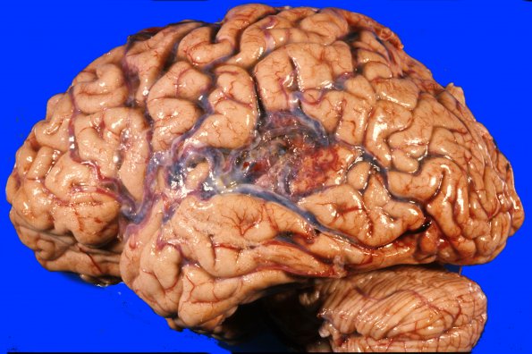
Case 1 History ---- The patient was a 65 year old man with AMML who developed pneumonia and sepsis in the presence of leukemic infiltrates in a variety of organs. He had no recognized symptoms of the AVM. ---- 1A1,2 The unfixed brain specimen shows an AVM in the temporal/parietal lobe. Many vessels of different sizes can be seen from this external view including multiple draining veins (arrows, 1A2).
 | Archive View |
Login
| Powered by Zenphoto