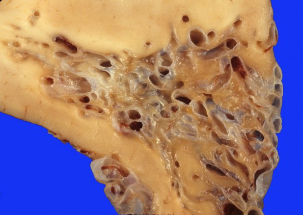
1A3,4 Higher magnification of the AVM in coronal section. There are numerous back to back thin- and thick-walled vessels as well as islands of orange discolored brain specimen.
 | Archive View |
Login
| Powered by Zenphoto