Table of Contents
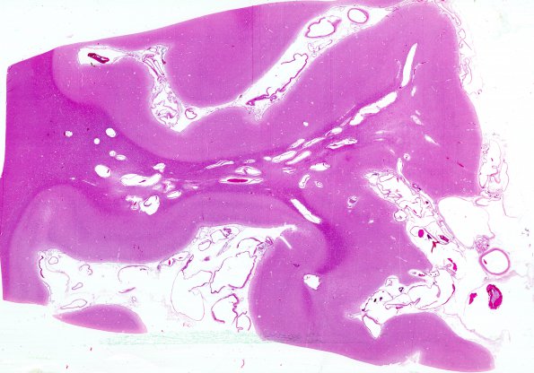
Washington University Experience | VASCULAR | AVM | 1C2 AVM (Case 1) H&E whole mount 3
However, the microscopic appearance shows thickened vasculature, smooth muscle and gliotic intervening parenchyma inconsistent with the diagnosis of venous angioma (H&E)

