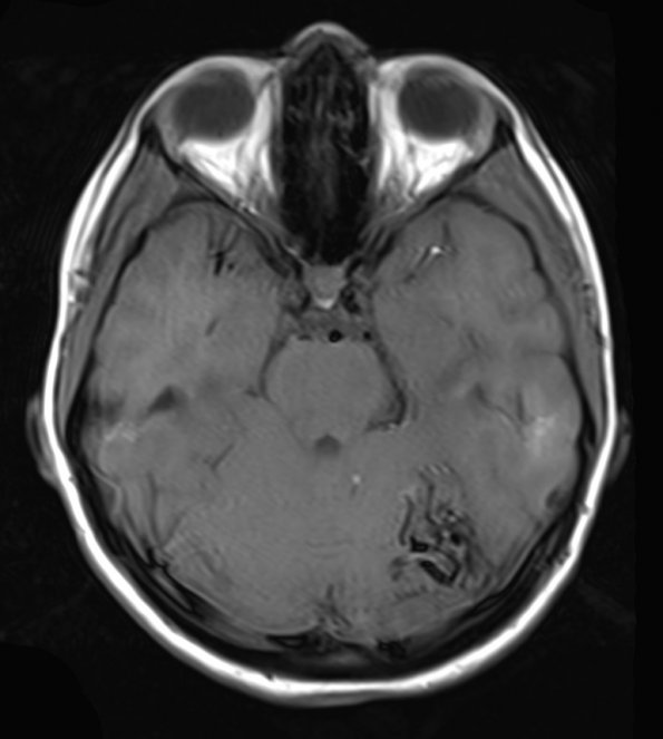
Case 20 History ---- The patient is a 17-year-old girl who presented with an especially severe 24 hour headache, blurred vision, and photophobia similar to a pattern of occipital and left sided migraine headaches she has experienced since age 8-9. In the emergency room, the patient's neurological examination was normal. However, subsequent imaging found a 3 cm tangle of flow-voids within the left superior cerebellar hemisphere consistent with an AVM. Onyx embolization resulted in 60%obliteration of the nidus. Five days later left suboccipital craniotomy was performed with resection of the AVM. ---- T1 weighted image with contrast (delayed?) of the AVM
 | Archive View |
Login
| Powered by Zenphoto