Table of Contents
Washington University Experience | VASCULAR | AVM | 25A1 AVM (Case 25) T2 W contrast 1 - Copy
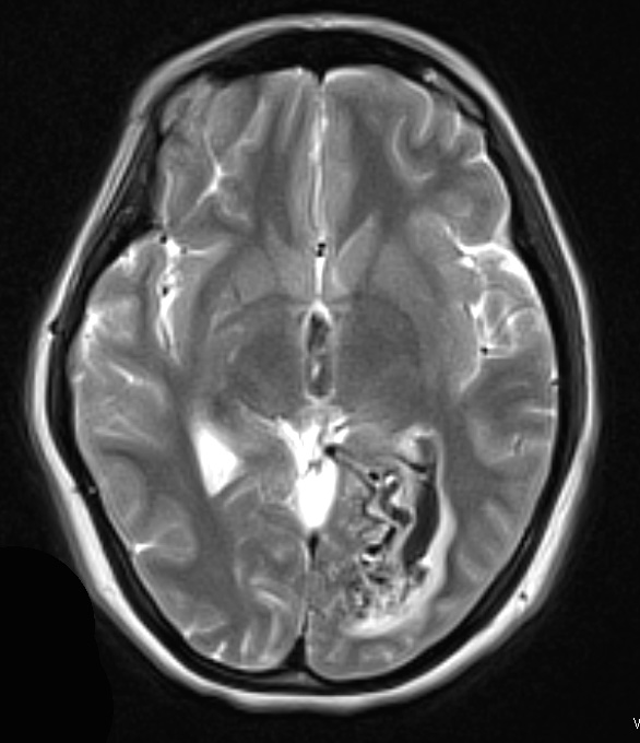
Case 25 History ---- The patient is a 34-year-old woman who presented with acute onset of headache. A head CT scan showed a left occipital intraparenchymal hematoma with intraventricular hemorrhage. Cerebral angiography showed a 1.9 cm left occipital AVM fed by the left PCA with drainage into vein of Galen/straight sinus and cortical venous drainage into the superior sagittal sinus. Pre-operative embolization with Onyx was performed one day before surgical resection. Operative procedure: Left occipital craniotomy for resection of AVM. ---- The AVM is demonstrated in this T2-weighted MRI image with contrast.

