Table of Contents
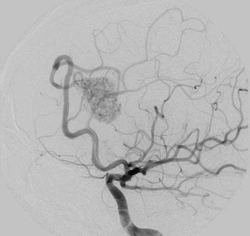
Washington University Experience | VASCULAR | AVM | 29A1 (Case 29) RICA PreEmboli angiogram - Copy
Case 29 History ---- This is a 36-year-old right-handed white female with a history of vertigo who had an incidental finding of a right frontal AVM in the absence of hemorrhage or seizures. Two weeks prior to presentation, she had an angiogram with embolization of the AVM with a good result. The AVM was 2.2 x 2.2 x 1.5 cm with right frontal location fed by the right anterior cerebral artery branches with the drainage to the superior sagittal sinus. The embolization with Onyx was successful with no residual filling of the AVM or early venous drainage. Two weeks later the AVM was resected ---- 29A1,2 Pre-(29A1) and post- (29A2) embolism angiograms from this patient.

