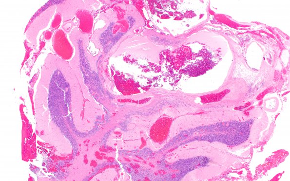
Routine H&E-stained sections of the resected right AVM demonstrate congeries of vessels of different sizes, wall thicknesses and composition with intercalated gliotic cerebellar parenchyma (H&E)
 | Archive View |
Login
| Powered by Zenphoto