Table of Contents
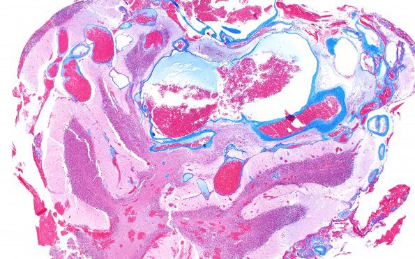
Washington University Experience | VASCULAR | AVM | 3B3 AVM (Case 3) A2 Trichrome 2X 2
3B3,4 A trichrome stain nearly sequential to those presented as image #3B1,2 shows disorganized vascular channels of different calibers and thicknesses separated by abnormal cerebellar parenchyma (Trichrome)

