Table of Contents
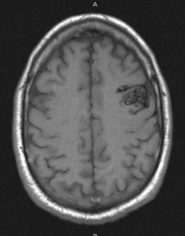
Washington University Experience | VASCULAR | AVM | 6A1 AVM (Case 6) T1 no contrast - Copy
Case 6 History ---- The patient is a 43 year old man with intractable depression for one year. CT and MRI examination and MR angiography showed a 2.0 x 2.7 cm AVM in the left frontal lobe, supplied by the frontal branch of the middle cerebral artery, with cortical venous drainage and mild to moderate stenosis of the largest draining vein. Operative procedure: Left craniotomy for resection of arteriovenous malformation ---- 6A1,2 Imaging techniques demonstrate a large AVM in T1-weighted (6A1) and T1-weighted with contrast administration (6A2).

