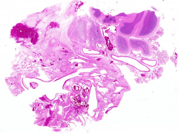
Whole mount of the cerebellum and AVM shows variation in vessel diameter and thickness with intercalated cerebellum ranging from areas with nearly complete neuron loss to normal appearing cerebellum in the right upper corner (H&E)
 | Archive View |
Login
| Powered by Zenphoto