Table of Contents
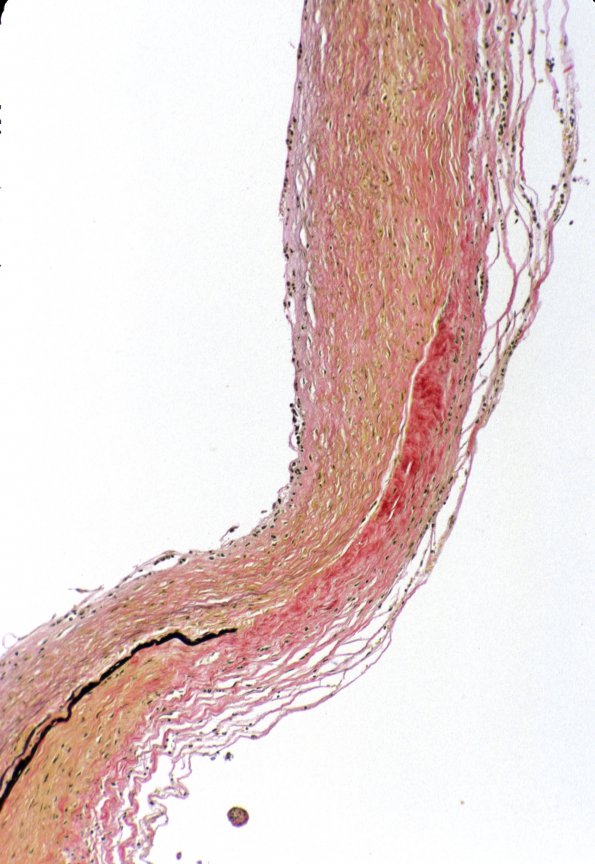
Washington University Experience | VASCULAR | Aneurysm - Saccular | 3B3 Aneurysm (Case 3) VVG 1
A close up shows the discontinuity and eventual loss of the internal elastic lamina at the origin of the aneurysm (VVG elastin stain) as well as marked wall thinning. At higher magnification there was an accumulation of neutrophils and scattered hemosiderin-laden macrophages at the site of interruption. (VVG)

