Table of Contents
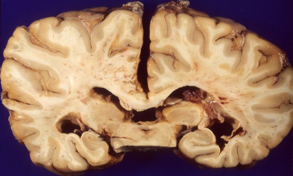
Washington University Experience | VASCULAR | Aneurysm - Saccular | 5A5 Aneurysm, saccular (Case 5) vertebral 5
In addition, there is a large area of subacute infarction involving the mesial aspect of the frontal, parietal, and occipital lobes in an anterior cerebral artery distribution. The cortex shows shrinkage and cystic tissue loss with underlying white matter softening and cystic tissue loss extending from the level of the splenium of the corpus callosum to its rostrum. ---- Sections of cerebral cortex diffusely show subarachnoid hemorrhage and hemosiderin-laden macrophages. The left parietal lobe lesion shows confluent areas of cystic tissue loss with numerous lipid-laden macrophages, inflammatory cells and surrounding astrocytic gliosis. All brainstem sections showed subarachnoid hemorrhage, hemosiderin and hematoidin-containing macrophages as well as spheroids in the midbrain/pons descending tracts. Focal Purkinje cell loss and astrocytosis was demonstrated in the cerebellar hemispheres.

